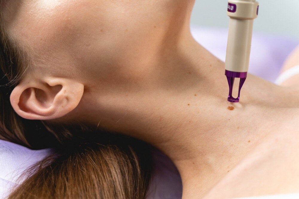
A mole that looks harmless may actually be a different type of growth, such as a seborrheic keratosis, wart, skin tag, dermoid cyst, or a lesion requiring urgent cancer evaluation. By the end, you will know which warning signs need prompt review, why dermoscopy and histopathology matter, and how removal methods differ in diagnostic value, scarring, healing, and recurrence risk.
Key takeaways
- Have every uncertain or changing mole assessed before removal.
- A biopsy helps identify lesions that could be skin cancer.
- Choose removal based on the lesion, not cosmetic preference alone.
- Confirm that removed tissue will be sent for laboratory analysis.
Which moles need assessment before removal?
Removal should wait until a clinician identifies the lesion. A skin assessment can distinguish a benign mole from seborrheic keratosis, wart, dermoid cyst, or skin tag; each has different treatment and diagnostic implications. The skin tag removal distinction matters clinically because destroying an uncertain pigmented lesion can delay melanoma diagnosis.
Dermoscopy, using magnified and illuminated examination, can reveal patterns hidden from normal view.
Use ABCDE and the ugly-duckling check as prompts for professional review, not as a diagnosis:
- Asymmetry: one half does not match the other.
- Border: the edge is ragged, blurred, notched, or uneven.
- Colour: the lesion contains several shades, including brown, black, red, blue, or white.
- Diameter: it measures over 6 mm, although melanoma can be smaller.
- Evolution: it changes in size, shape, colour, texture, or symptoms.
- Ugly duckling: it looks unlike your other moles, even without every ABCDE sign.
- Bleeding, crusting, persistent itching, pain, or a new changing lesion in adulthood also warrants review.
| Finding | Why it matters | Next step |
|---|---|---|
| Stable, similar-looking mole | Often fits a benign pattern | Arrange routine assessment if removal is wanted |
| Changing or symptomatic lesion | Change can signal malignancy | Prompt dermatology review and possible biopsy |
| Atypical or ugly-duckling lesion | Visual inspection cannot settle its identity | Dermoscopy; preserve tissue for histopathology |
Self-checking helps you notice change but cannot replace examination, photograph comparison, or biopsy when melanoma is suspected.
How a clinician examines a mole and decides on biopsy
A clinician begins by asking whether the mole is new, changing, bleeding, crusting, itchy, or painful. They also record past skin cancers, sun exposure, immune conditions, previous biopsies, and family history of melanoma. These details alter how urgently the lesion needs investigation.
The examination includes:
- Inspection in normal light, using the ABCDE check: Asymmetry, irregular Border, uneven Colour, Diameter over 6 mm, and Evolution. These are warning prompts, not a diagnosis; melanoma can be smaller than 6 mm.
- A search for the ugly-duckling sign: one lesion that looks unlike your other moles deserves attention, even without every ABCDE feature.
- Examination of the entire skin surface, scalp, nails, and nearby lymph-node areas when appropriate. This helps distinguish a mole from seborrhoeic keratosis, skin tag, wart, cyst, or a lesion needing urgent cancer assessment.
| Assessment method | What it reveals | What happens next |
|---|---|---|
| Dermoscopy | Magnified, illuminated pigment and vessel patterns hidden from the naked eye | The clinician decides whether monitoring, removal, or sampling is appropriate |
| Photograph comparison | Enlargement, colour, border, or shape changes over time | Evolution strengthens the case for biopsy |
| Biopsy for mole evaluation | Cells for microscopic examination | Histopathology confirms or excludes melanoma and other diagnoses |
If melanoma is possible, biopsy is safer than laser, cryotherapy, or cautery because destruction removes the evidence needed for histopathology. An excision biopsy with a narrow clinical margin, typically 2 mm, preserves the lesion for examination; complete excision may be chosen when a small uncertain mole can be removed and closed directly.
Which mole removal method fits the lesion?
A raised, clearly benign-appearing mole may suit a shave; a changing or pigmented lesion needs a method that preserves diagnostic tissue. Depth, recurrence risk, scarring, and pathology value matter as much as speed.
| Method | Depth and diagnostic value | Healing, scarring, recurrence, and best fit |
|---|---|---|
| Shave excision | Cuts across or just below the surface; may leave deeper melanocytic cells and provides limited assessment | Often heals without sutures, with a flat scar; residual cells can cause recurrent pigment. Selected raised, benign-appearing lesions only |
| Complete surgical excision with sutures | Removes the full depth and supplies an intact specimen for histopathology | Linear scar, local anaesthetic, stitches, and longer healing; lowest risk of leaving the lesion behind. Useful when diagnosis is uncertain |
| Punch excision | Removes a full-thickness circular core; provides strong diagnostic value for small lesions | Usually needs closure and leaves a small round scar; suitable for small pigmented or raised lesions |
| Elliptical excision | Removes the lesion deeply with an ellipse of surrounding skin; provides a complete specimen | Requires sutures and leaves a longer linear scar; useful when a wider margin or easier closure is needed |
| Electrosurgery | Burns tissue and usually leaves little or no specimen for pathology | Quick healing but possible pigment change, scarring, and recurrence; reserved for selected superficial, nonpigmented lesions |
| Cryotherapy | Freezes and destroys tissue without a specimen | Blistering, pigment change, and recurrence can occur; inappropriate for a pigmented or diagnostically uncertain mole |
| Laser treatment | Vaporises or alters tissue, usually preventing reliable pathology | Often heals quickly but pigment can recur and later recognition becomes harder; not a default for pigmented lesions |
If a mole regrows after shave removal, residual cells can create recurrent-nevus pigment in or around the scar, resembling melanoma. Ask how tissue will reach pathology before choosing any destructive method; never substitute acids, wart treatments, or home cutting.
What happens to removed tissue after the procedure?
Removed tissue is sent for histopathology, where a pathologist examines it under a microscope to identify the lesion and check whether abnormal cells are present. A pathology report can establish whether it is a benign nevus, atypical nevus, or melanoma; for melanoma, it records melanoma tumour thickness and other features that guide treatment.
Laser, cryotherapy, or cautery can destroy the specimen and leave a diagnostic gap.
- Ask when the pathology report is expected. Many clinics receive results within several days to two weeks, but confirm the timeline, who will explain it, and how you will receive the report.
- Follow up promptly if the report describes atypical, severely atypical, or malignant cells. You may need a wider excision, specialist referral, additional skin examination, or monitoring based on the diagnosis and margins.
- Keep the report and before-and-after photographs. If pigment returns after a shave removal, the recurrent nevus phenomenon can mimic melanoma in a scar, so the original findings help the next clinician interpret it.
Removal can cause a permanent scar, bleeding, infection, wound separation, altered sensation, pigment change, or delayed healing. Residual cells can also produce recurrence; a changing or bleeding regrowth needs assessment rather than another cosmetic treatment. “Scarless” and “no-downtime” claims do not remove these risks.
Questions to ask before booking mole removal
Before booking, ask who will assess the lesion, not just which device will remove it. A dermatologist should review its history, inspect the entire mole and surrounding skin, and use dermoscopy when appropriate. Ask whether a dermatologic surgeon will remove it and whether the tissue will be sent for histopathology.
- “Could this be something other than a benign mole, and do I need a biopsy?”
- “What findings would require a cancer specialist referral?”
- “Will you examine the rest of my skin and compare earlier photographs?”
- “What are the laser mole removal risks for this lesion, including recurrence and loss of a pathology specimen?”
- “What scar, pigment change, infection, bleeding, or wound problems fit my skin type and the treatment site?”
- “What should I do if it changes, bleeds, crusts, itches, or remains after removal?”
Use this comparison to test attractive promises:
| Claim | What to verify | Why it matters |
|---|---|---|
| Laser removal | Confirm the diagnosis and ask how pathology will be obtained | Laser can destroy suspicious tissue and leave no intact specimen |
| “No scars” | Ask for the expected scar outcome for your lesion, site, closure, and scar history | Any removal can leave a permanent or widened scar |
| “No downtime” | Request exact wound-care, activity, dressing, and return-to-work instructions | Quick recovery does not mean zero healing or zero risk |
At Khatri Clinic - Best Clinic in Camp 08048047677, ask the clinician to explain why the chosen method fits the diagnosis rather than accepting laser as the default. Never use wart acid, cutting tools, or home remedies on a changing or bleeding mole.
Frequently asked questions
Which moles need assessment before removal?
Have a clinician assess a mole before removal if it changes in size, shape, colour, or texture, bleeds, itches, hurts, has an irregular border, or differs from your other moles. Assessment also matters when the lesion could be a seborrheic keratosis, wart, dermoid cyst, or skin tag.
How does a clinician decide whether a mole needs a biopsy?
The clinician examines the lesion’s symmetry, border, colour, diameter, evolution, surface, and surrounding skin. A biopsy is chosen when the examination cannot confidently rule out melanoma or another condition requiring laboratory diagnosis.
Which mole removal method fits the lesion?
Excision removes the lesion with a margin and suits lesions that need complete removal or diagnosis. Shave removal removes a raised lesion close to the skin surface. Destructive treatments such as laser or cautery are unsuitable when tissue must be examined or melanoma has not been excluded.
What happens to tissue removed during mole removal?
When diagnosis matters, the clinician places the tissue in a labelled specimen container and sends it to a pathology laboratory. The pathology report identifies the lesion and confirms whether additional treatment is needed.
What should you ask before booking mole removal?
Ask who will examine the lesion, whether a biopsy is needed, which method will be used, whether the tissue goes to pathology, what scar to expect, and how you will receive the result.